
-
Mail us:
editor@raftpubs.org
Indexing & Abstracting
Full Text
Case ReportDOI Number : 10.36811/rjnnd.2022.110006Article Views : 0Article Downloads : 2
Unilateral tinnitus and sudden neurosensorial hearing loss caused by a looped vessel: An MRI case report
Abdulwahab Alahmari
Radiology Specialist, Radiology Department, Al-Namas General Hospital, Ministry of Health, Al-Namas City, Saudi Arabia
*Corresponding Author: Abdulwahab Alahmari, Radiology Specialist, Radiology Department, Al-Namas General Hospital, Ministry of Health, Al-Namas City, Saudi Arabia, Tel: +966562428716; Email: afaa99@hotmail.co.uk
Article Information
Aritcle Type: Case Report
Citation: Abdulwahab Alahmari. 2022. Unilateral tinnitus and sudden neurosensorial hearing loss caused by a looped vessel: An MRI case report. Res J Neuro N Disord. 4: 08-12.
Copyright: This is an open-access article distributed under the terms of the Creative Commons Attribution License, which permits unrestricted use, distribution, and reproduction in any medium, provided the original author and source are credited. Copyright © 2022; Abdulwahab Alahmari
Publication history:
Received date: 16 May, 2022Accepted date: 28 May, 2022
Published date: 31 May, 2022
Abstract
This is a case of neurovascular compression syndrome that lead to a sudden hearing loss on unilateral side in a female patient. The blood vessel is looping the vestibulocochlear nerve on the effected side by tinnitus and sudden neurosensorial hearing loss. All the theories and hypotheses about what could cause such symptoms will be discussed in this paper in details. The condition of the looping artery will be presented on an MRI scan T2 3D constructive interference in steady state images which is the best in visualizing conditions like this one.
Keywords: MRI; Tinnitus; Neurosensorial Hearing Loss; Neurovascular Compression Syndrome; Cerebellopontine Angle
Case report
A forty-six-year-old female patient came to the emergency room (ER) complaining of loss hearing on the left side and hearing buzzing sounds (pulsation) coming from inside her body (i.e. tinnitus). An MRI was done which revealed a looped vessel looping the vestibulocochlear nerve at the cerebellopontine angle (CPA). The patient was referred to the neurosurgery clinic for follow up and treatment. The findings are as the following; the 7th and 8th cranial nerves are intact and normal appearance of internal auditory canals. No CPA lesions and normal inner ear structures at both sides. Noted partial opacification of left mastoid air cells by mucosal thickening; however; preserved trabecular pattern. A Looped vessel (artery) could be seen crossing around the left vestibulocochlear nerve at the entrance of the left internal auditory meatus; however; no definite nerve kinking could be detected see (Figure 1-4).
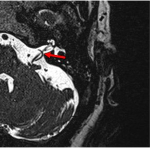
Figure 1: A brain MRI T2 3D constructive interference in steady state (CISS) shows the blood vessel (i.e. artery at the red arrow) looped around the vestibulocochlear nerve on the left side.
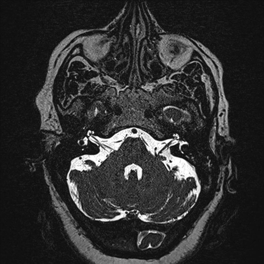
Figure 2: A brain MRI T2 3D CISS shows the blood vessel (i.e. artery) looped around the vestibulocochlear nerve on the left side.

Figure 3: A brain MRI T2 3D CISS shows the blood vessel (i.e. artery) looped around the vestibulocochlear nerve on the left side.

Figure 4: A brain MRI T2 3D CISS shows the blood vessel (i.e. artery) looped around the vestibulocochlear nerve on the left side.
Discussion
The artery that is looping the vestibulocochlear nerve is most likely to be a branch of the anteroinferior cerebellar artery (AICA) like another published case with similar findings [1]. A paper claimed that there is an anatomical relation between the loop and the vestibulocochlear nerve, but there is no relation between the loop and tinnitus [2]. In this case, there is no seen compression see (Fig. 5), but the artery is moving due to pulsations which means it can touch the nerve. There are other findings that might be involved −in this case− which are the mucosa has some thickening and mastoid air cells have opacification. As well, there is no statistical significance in between the loops of all types and tinnitus [2]. Furthermore, the angulation of the eighth nerve has no statistical significance [2]. But the author is asking the wrong questions. How many normal participants who have looped AICA of all types and have no symptoms of tinnitus or hearing loss? This is will rule out any involvement of neurovascular compression syndrome in cases like this case. The author refuted two hypotheses which are; the pulsatile compression and hypoperfusion both due to neurovascular compression without providing any evidence or claim! The issue is the author is citing paper that does not support the author’s claims! McDermott’s paper which the author citied basically found that loop type 2 and 3 are associated with hearing loss, but the size of the artery diameter has no statistical significance [3]. The MRI T2 3D CISS is the best in visualizing cranial nerves pathologies.

Figure 5. Illustration of the anatomical relation between AICA loop and vestibulocochlear nerve (CN VII).
There are three types of AICA loop; 1- lying in the CPA without entering the auditory canal, 2- entering the internal auditory canal, but not extending >50% of the internal auditory canal length, and 3- entering the internal auditory canal and extending >50% of the internal auditory canal. In this case, it fits with type 2 which the loop of the AICA reaches the entrance of the internal auditory canal without extending more than 50% of the canal length. Alahmari paper about dolichoectasia and neurovascular compression set the rules for classifying and making a distinction between pathological and normal anatomical variations [4-7]. The papers addresses when tortuousness and elongated arteries can cause symptoms or even death in cases like basilar artery occlusion due to dolichoectasia.
Conclusion
Unilateral tinnitus and sudden neurosensorial hearing loss can be caused by looping of the AICA on the eight cranial nerve. It might sound inductive to claim that looped vessel on 8th CN caused tinnitus, but there are many cases found the same findings. Therefore; from many findings we drew the conclusion. This case showed a looped vessel on the left side associated with hearing loss and tinnitus, while on the right side the artery is not looped and there is no hearing loss or tinnitus on the right side. Even if there is no seen compression, the pulsation and hypoperfusion will make the artery touch the nerve in a repeated fashion. Other factors were found to be have no statistical significance like; the type of the loop, the angulation of the 8th nerve, and the size of the artery diameter.
References
1. Ramly NA, Roslenda AR, Suraya A. 2014. Vascular loop in the cerebellopontine angle causing pulsatile tinnitus and headache: a case report. EXCLI Journal. 13: 192. Ref.: https://pubmed.ncbi.nlm.nih.gov/26417253/
2. Gultekin S, Celik H, Akpek S. 2008. Vascular loops at the cerebellopontine angle: is there a correlation with tinnitus? American Journal of Neuroradiology. 29: 1746-1749. Ref.: https://pubmed.ncbi.nlm.nih.gov/18653684/ DOI: https://doi.org/10.3174/ajnr.a1212
3. Mcdermott AL, Dutt SN, Irving RM. 2003. Anterior inferior cerebellar artery syndrome: fact or fiction. Clinical Otolaryngology & Allied Sciences. 28: 75- 80. Ref.: https://pubmed.ncbi.nlm.nih.gov/12680822/ DOI: https://doi.org/10.1046/j.1365- 2273.2003.00662.x
4. Alahmari A. 2020. Giant curved calcified basilar artery causing headache: A case report. Clinical Research in Neurology. 3: 1-4.
5. Alahmari A. 2021. Basilar Artery Occlusion: A Clinical Radiology Image. Austin J Radiol. 8.
6. Abdulwahab A. 2021. Basilar neuralgia vs. dolichoectasia: A report of three cases. O J Radio Med Img. 4: 1-9.
7. Alahmari A. 2020. A Second Case of Giant Atherosclerotic Basilar Artery Causing Headache: A Case Report.


