
-
Mail us:
editor@raftpubs.org
Indexing & Abstracting
Full Text
Case ReportDOI Number : 10.36811/osjs.2019.110002Article Views : 2577Article Downloads : 41
Mucosal stripping and muscular layer quilting in the treatment of anal canal duplication: Operative technique
Dogus Güney1* and Emrah Senel2
1Ankara Children’s Hematology Oncology Training and Research Hospital, Pediatric Surgery Clinic, Turkey
2Ankara Yildirim Beyazit University, Pediatric Surgery Clinic, Turkey
*Corresponding author: Dogus Güney, MD, Ankara Children’s Hematology Oncology Training and Research Hospital, Pediatric Surgery Clinic, Turkey, Tel: +90530 777 22 85; Fax: +90312 347 23 30: Email: dous_caliskan@hotmail.com
Article Information
Aritcle Type: Case Report
Citation: Dogus Güney, Emrah Senel. 2019. Mucosal stripping and muscular layer quilting in the treatment of anal canal duplication: Operative technique. Open Sci J Surg. 1: 04-07.
Copyright: This is an open-access article distributed under the terms of the Creative Commons Attribution License, which permits unrestricted use, distribution, and reproduction in any medium, provided the original author and source are credited. Copyright © 2019; Dogus Güney
Publication history:
Received date: 21 February, 2019Accepted date: 26 February, 2019
Published date: 28 February, 2019
Introduction
Anal canal duplication (ACD) is the least frequent digestive duplication [1]. It presents as a perineal orifice that ends with a tract along the anal canal [2]. Symptoms are often absent but can occur or worsen with age. Surgical excision of the duplicated anus is required because of the risk of infection and malignancy [3]. Here we present a patient with ACD that was treated with a technique that we pioneered called mucosal stripping and muscular layer quilting (MSMQ). This technique was performed for the first time in our clinic 11 years ago on 2 patients and has been described in a case report [4]. We describe the surgical technique here using a case presentation with photographs that illustrate the technique.
Case Report
A 10-month-old girl was brought to our pediatric surgery clinic after her parents noticed that there was a second anal opening. The opening was located just posterior to her normal anus Figure1. She had no history of infections in the area, no passage of stool from the duplicate anus, and no constipation or diarrhea. She was developing normally without any other medical problems or other identified anomalies. The patient was examined under general anesthesia. Exploration with a Hegar dilator revealed a blind-ending pouch with a depth of about two centimeters that was posterior to the normal anus but within the sphincter complex Figure2.
The duplicated anal canal was exposed with four hanging sutures. Normal saline solution was injected submucosally to promote hemostasis and facilitate mucosal dissection Figure 3. A mucosal incision was made, and the mucosa was stripped from the underlying muscle as in the Soave operation. The mucosa was removed as a whole Figure 4. The muscle layer of the duplicated anal canal was quilted with absorbable sutures. Perinoplasty was completed at the end of the operation Figure 5. The patient’s postoperative course was uneventful, and she was discharged home the second day after the surgery. At the 6-month follow-up exam, the patient had healed and had no further problems. The histological findings were consistent with an ACD diagnosis, showing stratified squamous epithelium with no transitional zone.
Figure: 1: Location of anal duplication.

Figure: 2: Exploration with a Hegar dilator.
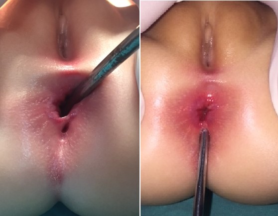
Figure: 3: Submucosal saline injection.

Figure: 4: Mucosal dissection.

Figure 5: Perinoplasty.

Discussion
ACD is an extremely rare pathology. In 2006, when we performed surgery for ACD on 2 patients at our clinic, there were 22 published cases of pediatric ACD [4]. The current literature shows a total 67 ACD cases, 6 in adults. Including the case presented here, 93% of ACD patients are female. ACD is most frequently (90%) a tubular anomaly without connections to the rectum and is usually asymptomatic [1,2,5]. If clinical symptoms are present, they can range widely from perianal pruritus, pain, fistula, abcess, constipation, stomach ache and vomiting to more vital ones like epidural abscess and sepsis. A detailed physical examination can identify ACD, and fistulography and MRI may also be helpful. There are 3 hypotheses in the literature concerning the origin of ACD, including an abnormal or duplicated cloaca or a recanalization failure [6,7]. In a study by Arakawa et al. of 28 human fetuses at 8–37 weeks of gestation, they observed no recanalization events. They hypothesized that an inferiorly extending anal sinus may result in an additional lumen for ACD [8].
Correction/removal of ACD is recommended because of the risk of infection and malignant changes, as reported by Dukes and Galvin [3]. Although we described the MSMQ technique 11 years ago, the literature shows that posterior sagittal/perineal techniques are still used for ACD treatment. Posterior sagittal/perineal techniques can lead to major complications, like permanent sphincter dysfunction, which require re-operation and can result in rectal injury, as well as to more minor complications like hematoma, infection, temporary bladder dysfunction, simple anal injury, and constipation [1,2,5]. The presence of associated pathologies, such as dermoid cyst, presacral tumor/abscess, lumbosacral meningocele, and spina bifida can change the prognosis and the surgical management [1,2]. However, most non-complicated cases of ACD cases should be treated using the MSMQ technique, which is simple and has a low risk of complications. In our 3 cases, we preferred to use the MSMQ technique to protect the sphincter mechanism against unnecessary dissection of the muscular layer and postoperative clinical outcomes were excellent. Since all reported malignant changes associated with ACD originated from the epithelium, we suggest that MSMQ should be the gold standard for the treatment of ACD.
In conclusion, the MSMQ technique is safe and easy to use. It could be the preferred first treatment option for non-complicated ACD.
References
- Van Biervliet, Maris E, Vande Velde S. 2013. Anal Canal Duplication in an 11-Year-Old-Child. Case Rep Gastrointest Med. [Ref.]
- Carpentier H, Maizlin I, Bliss D. 2009. Anal canal duplication: case reviews and summary of the world literature. Pediatr Surg Int. 25 :911-916.[Ref.]
- Dukes CE, Galvin C. 1956. Colloid carcinoma arising within fistulae in the anorectal region. Ann R Coll Surg Engl. 18 :246-261. [Ref.]
- Tiryaki T, Senel E, Atayurt H. 2006. Anal canal duplication in children: a new technique. Pediatr Surg Int. 22:560-1. [Ref.]
- Koga H, Okazaki T, Kato Y, et al. 2010. Anal canal duplication: experience at a single institution and literature review. Pediatr Surg Int. 26 :985-988. [Ref.]
- Van der Putte SC. 1986. Normal and abnormal development of the anorectum. J Pediatr Surg 21 :434-440. [Ref.]
- Nievelstein RA, Van der Werff JF, Verbeek FJ. 1998. Normal and abnormal embryonic development of the anorectum in human embryos. Teratology. 57 :70-8. [Ref.]
- Arakawa T, Eun Hwang S, Hyun Kim J. 2016. Fetal growth of the anal sinus and sphincters, especially in relation to anal anomalies. Int J Colorectal Dis. 31 :493-502. [Ref.]


