
-
Mail us:
editor@raftpubs.org
- ISSN : 2583-1534
Indexing & Abstracting
Full Text
Case ReportDOI Number : 10.36811/ojrmi.2019.110005Article Views : 19Article Downloads : 22
Visual failure following an invasive macroadenoma of the pituitary gland: A case report
Abdulwahab Alahmari
Radiology Specialist, Ministry of Health, Abha, Kingdom of Saudi Arabia
Corresponding Author: Abdulwahab Alahmari, Radiology Specialist, Ministry of Health, Abha, Kingdom of Saudi Arabia, Email: afaa99@hotmail.co.uk
Article Information
Aritcle Type: Case Report
Citation: Abdulwahab Alahmari. 2019. Visual failure following an invasive macroadenoma of the pituitary gland: A case report. O J Radio Med Img. 2: 30-32.
Copyright: This is an open-access article distributed under the terms of the Creative Commons Attribution License, which permits unrestricted use, distribution, and reproduction in any medium, provided the original author and source are credited. Copyright © 2019; Abdulwahab Alahmari
Publication history:
Received date: 26 November, 2019Accepted date: 06 December, 2019
Published date: 09 December, 2019
Abstract
A macroadenoma is a common disease with one type of invasion. In this case, a visual failure in both eyes followed a severe multiple invasion of macroadenoma which has all three types of invasion (sphenoid sinus, suprasellar, and cavernous sinus invasions).
Introduction
Pituitary tumors can invade and compress the surrounding structure. There are different types of invasion: sphenoid sinuses invasion is when a tumor breaks through the bone of the base of the sella turcica and crosses to the sphenoidal sinuses. The suprasellar invasion is when a tumor grows to cross the suprasellar region to the lateral or third ventricles. The cavernous sinus invasion is when a tumor invades the cavernous sinus which appears by surrounding the internal carotid artery (ICA) which passes in the cavernous sinus (cavernous sinus segment of the ICA). It required multiple macroadenoma patients to show different types of invasion in a paper [1]. In this case, all types of invasion are presented in a single case.
Case Report
A sixteen-year-old female patient came to the King Abdullah Medical City (neurology center) with right eye distorted vision which was very close to blindness (light can be seen, but the fuzziness causes objects cannot be determined) and a total left eye visual failure. The Goldman perimeter of the visual field test confirmed a complete visual dysfunction with a 0/20 visual acuity. The patient did not feel any menstrual issues or hormonal imbalance or symptoms except experiencing chronic headaches every day. In the left eye, the patient lost her vision suddenly and in the right eye, lost her vision, but in a fast-gradual rhythm. The patient let her sickness without a medical consultation from a Doctor for one year before she came to the neurology center. An MRI scan was performed for the patient which showed a large tumor in the pituitary gland > 10 mm which excluded microadenomas. The tumor’s dimensions are (18.6 cm is the width, 17.2 cm is the height, and 7.5 cm is the length). An MRI scan on time 1 (T1) pulse sequence with contrast media was preformed which showed all three types of invasion: 1- sphenoid sinus invasion, 2- suprasellar invasion, which affect the 3rd ventricle and lateral ventricles, and 3- cavernous sinus invasion, which affect the cavernous segment of the ICA (see Fig. 1 & 2). A biopsy and blood test were requested to identify the tumor functionality wither is it benign or malignant. The blood test came normal for the levels of the hormones and the biopsy confirmed the case as a benign macroadenoma and excluded pituitary carcinoma. Furthermore, an immunohistochemistry test confirmed atypical macroadenoma. The optic chiasma and optic nerves were compressed by the macroadenoma which caused the visual failure. The Neurologist decided to perform an endoscopic trans-sphenoidal surgery which used a 1 cm endoscope to remove the tumor. The endoscope was entered from the nose to cross the sphenoid bone to the pituitary gland. During surgery, all the judgments on the medical imaging study were confirmed. After eight-hour-long surgery, the patient woke up the next day and clinical exams were performed to check the patient’s visual ability. She can see 20/20 for both eyes after the tumor has been removed. The tumor was occupying on the left side more than the right side which caused the loss of vision suddenly on the left side.
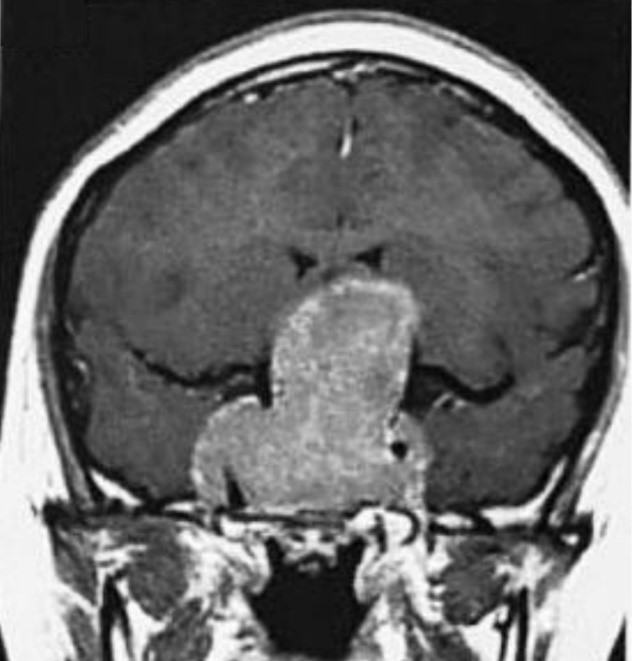
Figure 1: (A sixteen-year-old female). A brain MRI scan on the coronal section of the T1 with contrast. The scan revealed a severe invasion in the third ventricle and compressed lateral ventricles, an invasion in the cavernous sinus and the cavernous segment of the left ICA and compression against the medial aspect of the parietal and temporal lobes.
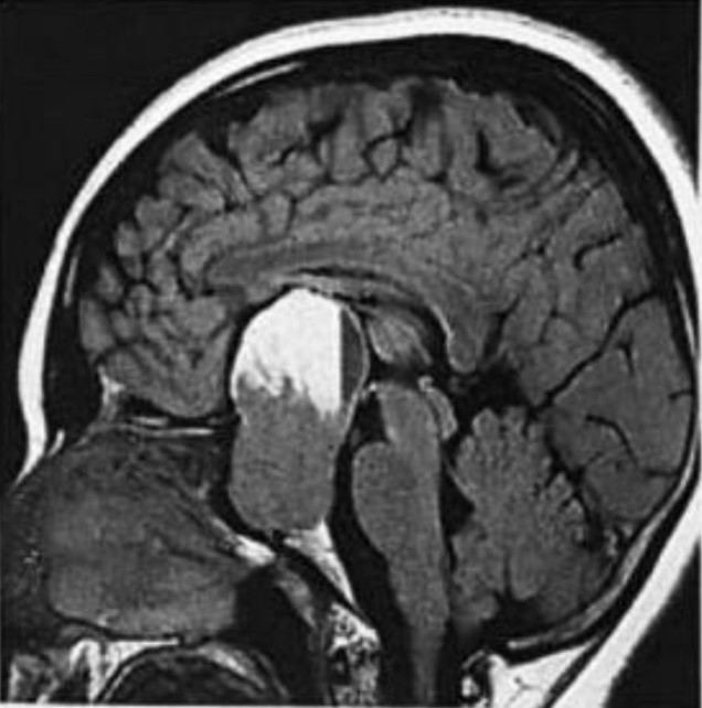
Figure 2: (A sixteen-year-old female). A brain MRI scan on the sagittal section of the T1 with contrast. The scan revealed a shenoidal invasion and compression of the frontal lobe from the posterior aspect. The tumor extends from the rostrum of the corpus callosum to the sphenoid bone.
Discussion
Visual failures and chiasmal dysfunctions are associated with pituitary macroadenomas [2]. All types of pituitary adenomas have the prevalence of 22.5% in imaging studies [3]. One of the common post-operative complications of endoscopic trans-sphenoidal surgery during removal of the pituitary macroadenoma is cerebrospinal fluid leakage [4].
References
1. Jia W, Lu R, Jia G, et al. 2013. Expression of pituitary tumor transforming gene (PTTG) in human pituitary macroadenomas. Tumor Biology. 34: 1559-1567. Ref.: https://www.ncbi.nlm.nih.gov/pubmed/23404407
2. Warnet A, Timsit J, Chanson P, et al. 1989. The effect of somatostatin analogue on chiasmal dysfunction from pituitary macroadenomas. Journal of neurosurgery. 71: 687-690. Ref.: http://tiny.cc/a7b8gz
3. Ezzat S, Asa SL, Couldwell WT, et al. 2004. The prevalence of pituitary adenomas: a systematic review. Cancer: Interdisciplinary International Journal of the American Cancer Society. 101: 613-619. Ref.: http://tiny.cc/19b8gz
4. Charalampaki P, Ayyad A, Kockro RA, et al. 2009. Surgical complications after endoscopic transsphenoidal pituitary surgery. Journal of Clinical Neuroscience. 16: 786-789. Ref.: https://www.ncbi.nlm.nih.gov/pubmed/19289287


