
-
Mail us:
editor@raftpubs.org
Indexing & Abstracting
Full Text
Letter to the EditorDOI Number : 10.36811/ojprm.2019.110002Article Views : 465Article Downloads : 14
A Broken Arrow: a rare complication of Endotracheal tube introducer
Shyh-Shyong Sim1*, Cheng-Hung How2 and Bo-Hwi Kang1
1Department of Emergency Medicine, Far Eastern Memorial Hospital, New Taipei City, Taiwan
2Department of Surgery, Far Eastern Memorial Hospital, New Taipei City, Taiwan
*Corresponding author: Shyh-Shyong Sim, MD, Department of Emergency Medicine, Far Eastern Memorial Hospital, No.21, Sec. 2, Nanya S. Rd., Banciao Dist., New Taipei City 220, Taiwan, Tel: 886-2- 8966-7000 Ext 1125; Email: vin_shyong@hotmail.com; vinshyong@gmail.com
Article Information
Aritcle Type: Letter to the Editor
Citation: Shyh-Shyong Sim, Cheng-Hung How, Bo-Hwi Kang. 2019. A Broken Arrow: a rare complication of Endotracheal tube introducer. Open J Pulm Respir Med. 1: 04-06.
Copyright: This is an open-access article distributed under the terms of the Creative Commons Attribution License, which permits unrestricted use, distribution, and reproduction in any medium, provided the original author and source are credited. Copyright © 2019; Shyh-Shyong Sim
Publication history:
Received date: 09 April, 2019Accepted date: 22 April, 2019
Published date: 24 April, 2019
Abstract
Complications from endotracheal tube introducer are rare and mostly involved mechanical trauma to airway structures. We report a rare complication while using endotracheal tube introducer during difficult airway management, which, we believed it was fragile after repeated sterilization.
Letter to the editor
The endotracheal tube introducer (ETI) is an inexpensive adjunct for managing difficult airway that is effective and easy to use. Nowadays, ETI is widely used by emergency physicians and anesthesiologists during endotracheal tube intubation. Complications from ETI are rare and mostly involved mechanical trauma to airway structures. By our own experience, we report a rare complication from ETI during difficult airway management.
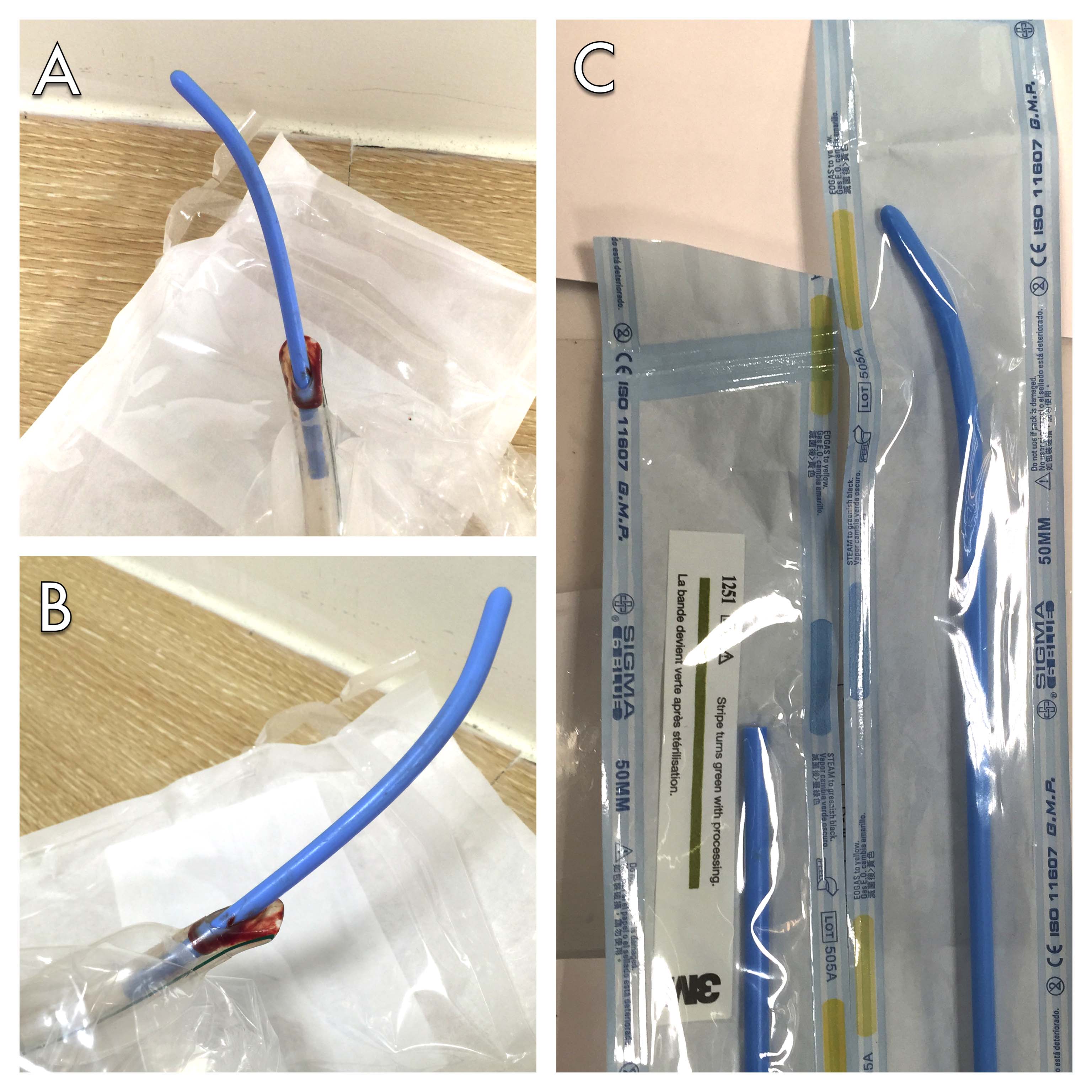
A 34-year-old male was brought to our emergency department in the early morning due to cardiac arrest. The patient was obese and vocal cord couldn’t be visualized under direct laryngoscopy. Several attempts of endotracheal tube intubation were unsuccessful. ETI was utilized and proper intubation was achieved after several trials. We confirmed the endotracheal tube placement by monitoring the number and waveform of end tidal CO2. After securing the airway, return of spontaneous circulation was achieved. However, the high airway pressure alarm kept going off from the ventilator. Bilateral breathing sound diminished and blood gas analysis showed CO2 retention. Suction tube met an obstacle while advancing through endotracheal tube at around 30cm. Fiberoptic bronchoscope was applied and noticed a blue, rounded and tube-like obstacle was stuck around the tip of endotracheal tube. We then replaced a new endotracheal tube. The obstacle was actually a broken tip of ETI stuck at the Murphy eye of the endotracheal tube (figure 1 A, B). Looking back, the first ETI was obviously shorter than a normal ETI (figure 1 C). After endotracheal tube replacement, patient’s airway pressure and bilateral breath sound became normal.
Figure: 1:
A and B, demonstration of the broken ETI tip stuck in the Murphy eye of endotracheal tube; C, the broken ETI (left side) is shorter than a normal ETI (right side).
The ETI, or commonly named as ‘bougie’, is now widely used as a convenient and inexpensive adjunct while managing airways. The ETI could be used with traditional laryngoscope, as an adjunct for video laryngoscopes and also blind digital intubation [1-3]. Complications from the ETI are rare and mostly involve mechanical trauma to airway structures [4]. Advancing the ETI might damage the larynx, trachea, or branches of the airway [5]. Most ETIs are single use, however, we believe that many medical departments reuse it after proper sterilization. Our experience demonstrated a rare complication that happened while using an ETI, which might become fragile after repeated sterilization. We strongly recommend a routine check for its completeness after using ETI. In the future, while physicians encounter high airway pressure and suspect an obstacle or foreign body in the airway after using ETI for intubation, a broken ETI should be considered and excluded.
Acknowledgments
We thank all the nursing and medical staff of the Department of Emergency Medicine, Far Eastern Memorial Hospital, New Taipei City, Taiwan.
Conflict of interest statement
The authors have no commercial associations or sources of support that might pose a conflict of interest.
References
- Takenaka I, Aoyama K, Iwagaki T, et al. 2009. Approach combining the airway scope and the bougie for minimizing movement of the cervical spine during endotracheal intubation. Anesthesiology. 110: 1335. [Ref.]
- Inoue H, Saito T, Kamishima K, et al. 2008. Successful nasal intubation using airway scope with gum elastic bougie in a case of difficult airway. Masui. 57: 457-459. [Ref.]
- Rich JM. 2008. Successful blind digital intubation with a bougie introducer in a patient with an unexpected difficult airway. Proc (Bayl Univ Med Cent). 21: 397-399. [Ref.]
- Kadry M, Popat M. 1999. Pharyngeal wall perforation--an unusual complication of blind intubation with a gum elastic bougie. Anaesthesia. 54: 404-405. [Ref.]
- Marson BA, Anderson E, Wilkes AR, et al. 2014. Bougie-related airway trauma: dangers of the hold-up sign. Anaesthesia. 69: 219-223. [Ref.]


