Indexing & Abstracting
Full Text
Case ReportDOI Number : 10.36811/ojgor.2020.110016Article Views : 3Article Downloads : 4
Bladder Stone: An Uncommon Cause of Urinary Incontinence
Ekwedigwe KC1, Isikhuemen ME2*, Lengmang S1,Sunday-Adeoye I1 and Eliboh MO1
1National Obstetric Fistula Centre, Abakaliki, Nigeria
2University of Benin Teaching Hospital, Edo State, Nigeria
*Corresponding Author: M.E. Isikhuemen, University of Benin Teaching Hospital, Edo State, Nigeria, Tel: +2348050638600; Email: maradona4real2002@yahoo.com
Article Information
Aritcle Type: Case Report
Citation: Ekwedigwe KC, Isikhuemen ME, Lengmang S, Sunday-Adeoye I, Eliboh MO. 2020. Bladder Stone: An Uncommon Cause of Urinary Incontinence. O J Gyencol Obset Res. 2: 30-33.
Copyright: This is an open-access article distributed under the terms of the Creative Commons Attribution License, which permits unrestricted use, distribution, and reproduction in any medium, provided the original author and source are credited. Copyright © 2020; Ekwedigwe KC
Publication history:
Received date: 30 April, 2020Accepted date: 06 May, 2020
Published date: 07 May, 2020
Abstract
Background: Bladder stones contribute about 5% to urinary tract stones. Bladder calculi is not a common cause of urinary incontinence and huge bladder stones are not common in routine clinical practice. Our aim, in this case, was to report a patient with huge bladder stone and share our experience in the management.
Case presentation: She is a 41-year-old grand multipara who presented to the National Obstetric Fistula Centre, Abakaliki with a history of urinary incontinence. Presence of bladder stone was felt with a metal urethral catheter during clinical examination and this was confirmed with imaging. She was managed with transvaginal cystolithotomy.
Conclusion: Huge bladder stone is uncommon but can be a cause of urinary incontinence. Transvaginal cystolithotomy is a safe approach to its management.
Keywords: Bladder stone; Urinary incontinence; Cystolithotomy
Introduction
Bladder stones contributes 5% of stones of the urinary tract [1]. It is more common in males than females [2]. About 5% of all bladder stones occur in women [3]. The short urethra in women facilitates easy passage of bladder stone [4]. Occurrence of bladder stones are usually associated with foreign bodies or urine stasis [3]. Women with bladder calculi may present with irritable symptoms, haematuria or recurrent infections though it can be asymptomatic [3]. A study done in Dubai showed that acute urinary retention was the most common modality of presentation in patients with bladder stone [5]. Bladder stones causing urinary incontinence have previously been reported in the literature [6]. If there is urinary incontinence following bladder stones, this may lead to psychosocial problems. Though there are predisposing factors to stone formation, some can occur without any underlying urinary tract pathology [7]. Endoscopic transurethral fragmentation is the treatment option for bladder stones, but when the stone is large, percutaneous endoscopic disintegration or open suprapubic cystolithotomy is the preferred option [3]. Transvaginal cystolithotomy is also safe [8]. We report a case of huge bladder stone in a grand multiparous woman and share our experience in its management.
Case report
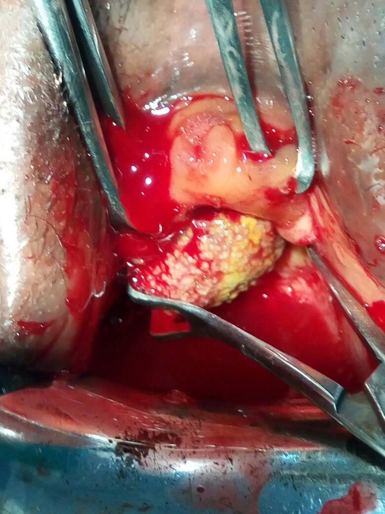
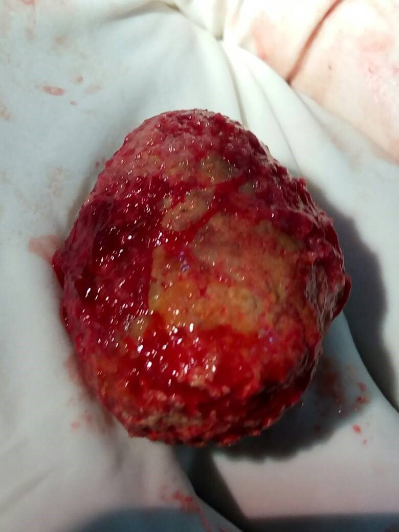
She is a 41-year-old woman with five children who presented with urinary incontinence of three years duration. There was a positive history of urgency and urge incontintnce. Symptoms worsened with time. She had subtotal hysterectomy following uterine rupture in her last delivery five years prior to presentation. She also gave a history of painful urination and urinary frequency. There was no history of diabetes or any other neurogenic disorder. There were no other significant findings in the history. On examination, she was in no obvious distress. Type 2 female genital mutilation was noted. Cough impulse was positive. A bladder stone was demonstrated in the vesicourethral region with a metal catheter. No urethrovaginal or vesicovaginal fistula was demonstrated. An assessment of urinary incontinence secondary to bladder stones was made. Basic investigations done were essentially normal and imaging confirmed the presence of urinary calculi. Patient was counselled on the diagnosis and scheduled to have transvaginal cystolithotomy (Figure 1). A stone measuring 10 X 8 X 6cm was removed from the bladder (Figure 2). She then had copious bladder irrigation with normal saline. The bladder incision was closed in layers with absorbable sutures. She was then catheterized. In the post operative period, she was managed with antibiotics and analgesics. Urethral catheter was left in-situ for 14 days. Post operative dye test done did not suggest any urethrovaginal or vesicovaginal fistula. She had a dry vulva and was discharged after removal of urethral catheter. She has remained without any urinary symptom on follow up.

Figure 1: This shows the process of removing the stone from the bladder.

Figure 2: This shows the stone after removal from the bladder.
Discussion
Huge bladder stone is not a common finding. It is not a usual cause of urinary incontinence in women. Women with bladder stones may present with haematuria, dysuria and urinary incontinence, though these features are not pathognomonic for bladder stones [4]. As shown in this case report, our patient had urinary incontinence associated with urgency. There was also associated history of dysuria and frequent urination. The symptoms of patients with bladder stones may depend on the location of the stone within the bladder. Stones that are close to the urethral opening are likely to cause obstructive symptoms compared to stones in other parts of the bladder.
There are risk factors for stone formation, although it may occur without any known predisposing factor [7]. The risk factor for stone formation is not clear in the index patient although she had five pregnancies which may predispose her to urinary tract stones. Elevated progesterone and mechanical compression by the foetus places pregnant women at risk of stone formation due to urinary stasis [9,10].
There are various management options for bladder stones. The patient presented in this case report had transvaginal cystolithotomy. This has been shown to be a safe procedure for removal of bladder stone [8]. In patients with small bladder stones and urethral problems, both pathologies can be treated endoscopically [4]. Endoscopic transurethral fragmentation of the stone is the preferred option in the management of bladder stones but when stone is large, percutaneous endoscopic disintegration or open cystolithotomy is useful [3,6]. Extracorporeal shockwave lithotripsy is simple, effective, and well tolerated [3]. Due to the fact that our patient had a large bladder stone, she had open surgery for removal of bladder stone. Open surgery for bladder stones has a success rate of about 100% [11].
Conclusion
This case report is of considerable interest because bladder stone of this size is uncommon in routine practice. Patients with bladder stones commonly have predisposing factors though this was not obvious in the index patient. Huge bladder stones can be safely managed using transvaginal cystolithotomy.
Consent
Informed consent was obtained from the patient that was presented in this case report.
References
1. Tahtali IN, Karatas T. 2014. Giant bladder stone: a case report and review of the literature. Turk J Urol. 40: 189-191. Ref.: https://www.ncbi.nlm.nih.gov/pmc/articles/PMC4548388/
2. Aydogdu O, Telli O, Burgu B, et al. 2011. Intravesical obstruction results as giant bladder calculi. Urol Assoc J. 5: 77-78.
3. Kobi S, Peter D. 2012. Urinary bladder stones in women. Obstet Gynecol Surv. 67: 715 -725. Ref.: https://bit.ly/2WqUNVe
4. Tasi Gk, Onuk O, Balci MBC, et al. 2014. Giant Bladder Stone in a Young Woman. JAREM. 4: 132-134. Ref.: https://bit.ly/2yAxPD0
5. Hammad FT, Kaya M, Kazim E. 2006. Bladder calculi: Did the clinical picture change? Urology. 67: 1154-1158. Ref.: https://bit.ly/3c79e7C
6. Basatac C, Cicek MC. 2016. An Uncommon Cause of Urge Urinary Incontinence in Female Patient: Huge Bladder Stone. SM J Urol. 2: 1015.
7. Bayindir M, Resorlu B, Sarikaya S, et al. 2012. Giant Bladder Stone In A Middle-Aged Female Patient without any Predisposing Factor: Case Report. Ankara Universitesi Tip Fakultesi Mecmuasi. 65: 179-182.
8. Hudson CO, Sinno AK, Northington GD, et al. 2014. Complete transvaginal surgical management of multiple bladder calculi and obstructed uterine procidentia. Female Pelvic Med Reconstr Surg. 20: 59-61. Ref.: https://bit.ly/3b85W2z
9. Semins MJ, Matlaga BR. 2013. Management of urolithiasis in pregnancy. Int J Womens health. 5: 599-604. Ref.: https://www.ncbi.nlm.nih.gov/pmc/articles/PMC3792830/
10. Smith CL, Kristensen C, Davis M, et al. 2001. An evaluation of the physiochemical risk for renal stone disease during pregnancy. Clin Nephrol. 55: 205-211. Ref.: https://bit.ly/3frHBYG
11. Telha KA, Alkohlany K, Alnono I. 2016. Extracorporeal shockwave lithotripsy monotherapy for treating patients with bladder stones. Arab J Urol. 14: 207-210. Ref.: https://bit.ly/2W94uIW


