Indexing & Abstracting
Full Text
Case ReportDOI Number : 10.36811/jcri.2019.110006Article Views : 15Article Downloads : 17
Role of genetic diagnosis in thyroid neoplasms
Vaktangova N*, Garcia Castañon S, Martinez Gago MD, Corrales B and Rodríguez Aguilar R
Garcia Moreira V. University Hospital of Cabueñes, Gijón. Spain
*Corresponding author: Vaktangova N, Garcia Moreira V. University Hospital of Cabueñes, Gijón. Spain. Email: nmedik1@gmail.com
Article Information
Aritcle Type: Case Report
Citation: Vaktangova N, Garcia Castañon S, Martinez Gago MD, et al. 2019. Role of genetic diagnosis in thyroid neoplasms. J Case Rept Img. 1: 40-42.
Copyright:This is an open-access article distributed under the terms of the Creative Commons Attribution License, which permits unrestricted use, distribution, and reproduction in any medium, provided the original author and source are credited. Copyright © 2019; Vaktangova N
Publication history:
Received date: 20 May, 2019Accepted date: 04 June, 2019
Published date: 06 June, 2019
Summary
Carcinoma medullary thyroid is a rare, malignant neoplasm. Affected patients can be hereditary carriers, but in most cases they are spontaneously affected. In hereditary cases, prophylactic surgery is recommended at an early age. We present a case of the patient carrier of mutation of the proto-oncogene type RET with medullary thyroid carcinoma prophylactic surgery already in adulthood.
Keywords: Medullary thyroid carcinoma; MEN 2; Prophylactic surgery
Introduction
Medullary thyroid carcinoma is a malignant [1,2] neoplasm that originates in the parafollicular cells of the thyroid gland (also called the C cells). It is a rare neoplasm, representing 5% of all thyroid cancers. It occurs spontaneously in 84% of cases or is hereditary in 16% of cases. In the cases of the hereditary1 form it can be in the context of familial medullary thyroid carcinoma or in the form of a multiple endocrine neoplasia type 2 (MEN 2), which is associated with different mutations of the protooncogene type RET in chromosome 10q11.2. The clinical markers in this case are calcitonin and carcinoembryonic antigen (CEA).
Clinical Case
Male patient, 54 years, with a family history of MEN 2, bilateral pheochromocytoma and medullary thyroid carcinoma attended the endocrinology review. At the time of examination, no goiter is palpated nor cervical adenopathies. The patient is given complementary tests. The blood test shows the following results: Hb 15.0 g / dl; Red blood cells 4.88 x 10E / μL; Platelets 244x1000 / μ ;, Leukocytes 7.43x1000 / μL; Neutrophils 61.2%; Lymphocytes 23.3%; Monocytes 9.4%; Creatinine 0.88 mg / dL; Albumin 42 g / L; Calcium 9.6 mg / dL; GPT 27U / L; GOT 20 U / L; GGT 36U / L; TSH 1.42 μUI / ml; PTH 67.5 pg / mL; Vitamin D 25 (OH) 13.8 ng / mL; Calcitonin 48pg / mL; CEA 1.6 ng / ml. Genetic study shows the presence of the alteration c.1853> A (p.Cys618Tyr) in the RET gene in heterozygosis in the sample studied.
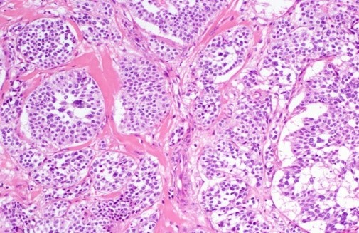
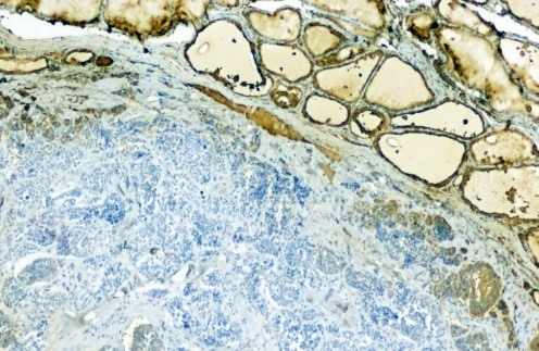
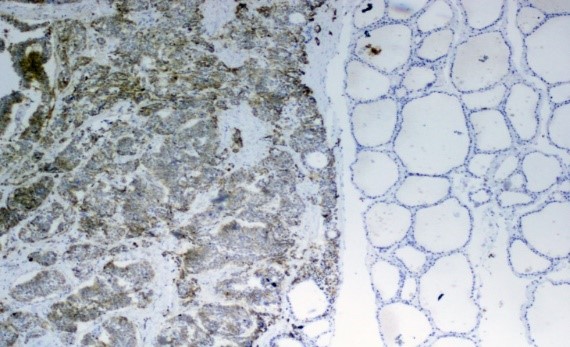
In thyroid ultrasound thyroid gland of normal size and eco-structure is appreciated. A poorly defined hypoechoic nodule is detected in the right thyroid lobe of size 0.56 x 0.58 x 0.66 cm. No adenopathies have been detected. The results of the ultrasound are confirmed in the cervical CT scan: Normal-sized thyroid gland, with a nodule in the right thyroid lobe measuring 5 mm in diameter. The patient underwent a total prophylactic thyroidectomy and right recurrent voiding. Pathological anatomy results describe: Medullary thyroid microcarcinoma, image 1, Hematoxylin-Eosin staining) of 0.5 cm in diameter in right hemithyroid and two foci of medullary thyroid microcarcinoma, 0.3 and 0.1 cm in diameter in left hemithyroids. No lymph node involvement is observed. Histological stains of Calcitonin, Thyroglobulin, CEA (carcinoembryonic antigen) and positive Chromogranin (images 2-5). In the postoperative analysis, the results were as follows: Albumin 40 g / L; Calcium 9.3 mg / dL; TSH 0.64 μUI / ml; Calcitonin 12.3 pg / mL; CEA 1.6 ng / ml.

Image 1: Medullary thyroid carcinoma. Staining: H & E x 100.

Image 2: Medullary thyroid carcinoma. Staining: Calcitonin x 20.

Image 3: Medullary thyroid carcinoma. Staining: Thyroglobulin x 100.

Image 4: Thyroid medullary carcinoma. Staining: CEA x 20.

Image 5: Thyroid medullary carcinoma. Staining: Chromogranin x 20.
Discussion
For the study of hereditary [1] thyroid cancer, it is recommended to perform a genetic study and measurement test of calcitonin stimulated with pentagastrin in people with a personal history of thyroid cancer, neuroendocrine neoplasms, history in first degree relatives [2]. In the presence of the RET gene mutation, it is suggested, supported by numerous studies, to perform a prophylactic surgery at an early age [3] to prevent the evolution from preneoplastic to neoplastic stage both in patients with antecedentes and in those who are asymptomatic. In the case of our patient, prophylactic surgery has been performed at an adult age. The patient is currently continuing with endocrinology reviews.
Conclusions
When it comes to patients with thyroid neoplasms it is very important to carry out the genetic study and provide all the corresponding information. In patients with known hereditary diseases, the genetic study becomes even more important. In the case of our patient, if the genetic study had been carried out at an early age, his prognosis would be more favorable.
References
- Dotto J, Nosé V. 2008. Familial thyroid carcinoma: a diagnostic algorithm. Adv Anat Pathol. 15: 332-349. [Ref.]
- Maia AL, Siqueira DR, Kulcsar M, et al. 2014. Diagnosis, treatment, and follow-up of medullary thyroid carcinoma: Recommendations by the Thyroid Department of the Brazilian Society of Endocrinology and Metabolism. Arq Bras Endocrinol Metab. 58: 667-700. [Ref.]
- Rosenthal MS, Diekema DS. 2011. Pediatric ethics guidelines for hereditary medullary thyroid cancer. International Journal of Pediatric Endocrinology. [Ref.]


