Indexing & Abstracting
Full Text
Case ReportDOI Number : 10.36811/gju.2021.110013Article Views : 105Article Downloads : 97
Inverted papilloma, a Crying wolf of the urinary bladder
Manjeet Kumar1*, Manika sharma2, Kirti Rana3, Kailash Barwal4 and Sanjeev Chauhan5
1Assistant Professor, Department of Urology, IGMC Shimla, Himachal Pradesh, India
2Senior Resident Department of Pathology, IGMC Shimla, Himachal Pradesh, India
3Senior Resident Department of Urology, IGMC Shimla, Himachal Pradesh, India
4,5Associate Professor, Department of Urology, IGMC Shimla, Himachal Pradesh, India
*Corresponding Author: Manjeet Kumar, Assistant Professor, Department of Urology, IGMC Shimla, Himachal Pradesh, India, Email: manjeetkumar.1014@gmail.com
Article Information
Aritcle Type: Case Report
Citation: Manjeet Kumar, Manika sharma, Kirti Rana, et al. 2021. Inverted papilloma, a Crying wolf of the urinary bladder. Glob J Urol. 3: 12-15.
Copyright: This is an open-access article distributed under the terms of the Creative Commons Attribution License, which permits unrestricted use, distribution, and reproduction in any medium, provided the original author and source are credited. Copyright © 2021; Manjeet Kumar
Publication history:
Received date: 15 September, 2021Accepted date: 22 September, 2021
Published date: 24 September, 2021
Abstract
Inverted papilloma is a benign, non-invasive and innoxious bladder tumour rarely seen. These tumours are associated with chronic urinary tract infection, bladder neck obstruction, and they appear similar to other bladder tumours. Inverted papilloma is diagnosed on histopathological examination. We report a case of inverted papilloma who presented and appeared as bladder tumour but on subsequent pathological examination diagnosed as inverted papilloma.
Keywords: Inverted papilloma; Benign; TCC (Transitional cell carcinoma); PUNLMP (Papillary urothelial neoplasm of low malignant Potential)
Introduction
Inverted papilloma is a benign proliferative lesion with unknown aetiology. It is found to be associated with chronic inflammation or bladder outlet obstruction, smoking, bladder neck obstructions. It can occur throughout the urinary bladder but the most common location is on the trigone. It accounts for less than 1% of all bladder tumours. Painless gross haematuria is the most common representing symptom; with microscopic haematuria and irritative voiding symptoms also common. [1,2] We describe a case report of a patient presented with haematuria, and on pathological examination found to be inverted papilloma.
Case summary
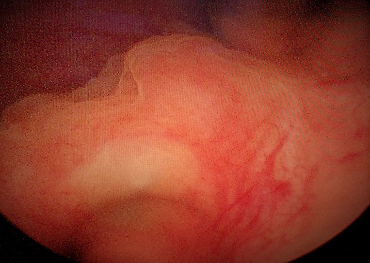
A 39 years male adult presented with a history of haematuria for 1 month. He did not have history of chronic urinary tract infections, stones in urinary tract and obstructive urinary tract symptoms; although he is active smoker. Blood investigations were Haemoglobin 13.5 gm%, TLC 5500, urea 22 mg%, creatinine 0.6mg%. Ultrasonography revealed a polypoidal urinary bladder lesion 2x 2 cm in the posterior wall of the urinary bladder near the bladder neck. Cystoscopy examination revealed a 2x2 cm tumour arising from the bladder neck. (Figure 1).

Figure 1: Cystoscopy shows raised, smooth, polypoidal urinary bladder lesion 2 x 2 cm on trigone of urinary bladder.
Transurethral resection of bladder tumour (TURBT) was done and resected tissue was sent for histopathological examination. Histopathogical examination was suggestive of inverted papilloma urinary bladder. Figure 2a,2b.

Figure 2: a,b. Tumor cells in inverted growth pattern in form cords and sheets from urothelium.
The postoperative period was uneventful and he was discharged from the hospital after removal of per urethral catheter on postoperative day 1. Histopathological examination revealed features of inverted papilloma with mild atypia and no tumour invasion. He is followed with cystoscopy and ultrasound examination at 3 months and one year of TURBT.
Discussion
Paschim (1927) first described inverted papilloma and named by Potts and Hirst (1963). Inverted papilloma comprises about 1-2% of all bladder neoplasms [3,4].
It behaves in a benign fashion with a less than 1% chance of recurrence. Previously Papilloma was considered to be a low-grade Ta malignant bladder tumour, nowadays they are considered benign and non-invasive tumours. Mutations of Fibroblast growth factor receptor -3(FGFR3) were seen in papilloma as well as bladder tumours, however, they lack markers of aggressive behaviour, including TP53 and RB mutations. [5]. It is diagnosed typically in males (M: F 7.3:1) in sixth to seventh decade of life. The most probable causes of inverted papilloma are smoking, chronic bladder infection, and urinary obstruction [6].
The most common symptoms reported are irritative urinary symptoms, gross haematuria, microscopic haematuria, suprapubic pain, etc. The most common location of inverted papilloma is bladder neck and trigone. Cumming et al purposed inverted papilloma as a hyperplastic lesion instead of a neoplastic lesion to chronic inflammation agents, or irritative agents. [7]. Inverted papilloma on cystoscopy is seen as a pedunculated, such as papillary, polypoid, and seaweed-like, or sessile mass with a smooth surface. They are typically 1-2 cm in size, but can sometimes be large size. Henderson et al described endophytic extension of uniform urothelial cells from urothelium (surface epithelium) as inverted papilloma [8]. Inverted papilloma pathological characteristics are growth of urothelium with inverted pattern, mature urothelium having smooth surface, uniform morphology of epithelium, Trabecular and nested (smooth) array of tumour cells, few or no mitotic figures, occasional non keratinising squamous metaplasia or microcysts, non-invasive and no exophytic components. [8,9]. Inverted papilloma can be confused with PUNLMP, cystitis cystica, transitional cell carcinoma, and Brun’s cell nests. Piozzi et al described inverted papilloma as a potential risk factor for urothelial cancer. He showed that 1% of patients developed urothelial carcinoma after surgery for inverted papilloma in approximately 27 months. So inverted papilloma should be followed with cystoscopy although adequate resection is sufficient treatment needed in most cases [2,9]. Complete resection is an adequate treatment for inverted papilloma. Recent studies suggest IPB as a benign neoplasm, however, it is mandatory to exclude associated invasive bladder tumours. These tumours are risk factors for invasive bladder tumours, so follow-up is mandatory although less stringent. Our case who was a young male with a 2x2 cm tumour was treated with complete resection. On follow up no recurrence was seen up to 2 years. We plan to follow him with yearly cystoscopy for 5 years.
Conclusions
Inverted Papilloma are uncommon benign bladder tumours. Accurate histopathological diagnosis is key as morphology is similar to invasive transitional cell carcinoma. Careful follow-up is necessary as they are at risk for invasive TCC.
Abbreviations
• PUNLMP papillary urothelial neoplasia of malignant potential
• TURBT Transurethral resection of bladder tumours
• TCC Transitional cell carcinoma of urinary bladder
References
1. Jones TD, Zhang S, Lopez-Beltran A, et al. 2007. Urothelial carcinoma with an inverted growth pattern can be distinguished from inverted papilloma by fluorescence in situ hybridization, immunohistochemistry, and morphologic analysis. Am J Surg Pathol. 31: 1861-1867. Ref.: https://pubmed.ncbi.nlm.nih.gov/18043040/ Doi: https://doi.org/10.1097/pas.0b013e318060cb9d
2. Picozzi S, Casellato S, Bozzini G, et al. 2013. Inverted papilloma of the bladder: a review and an analysis of the recent literature of 365 patients. Urol Oncol. 31: 1584-1590. Ref.: https://pubmed.ncbi.nlm.nih.gov/22520573/ Doi.: https://doi.org/10.1016/j.urolonc.2012.03.009
3. Paschkis R, Adenome der Harnblase U. Ztschr, et al. 1927. 21: 315-325.
4. Potts IF, Hirst E. 1963. Inverted papilloma of the bladder. J Urol. 90: 175-179. Ref.: https://pubmed.ncbi.nlm.nih.gov/14044188/ Doi: https://doi.org/10.1016/s0022-5347(17)64384-2
5. Liang Cheng, Antonio Lopez-Beltran, David G, et al. 2012. Bladder Pathology.
6. Guo A, Liu A, Teng X. 2016. The pathology of urinary bladder lesions with an inverted pattern. Chin J Cancer Res. 28: 107-121. Ref.: https://pubmed.ncbi.nlm.nih.gov/27041933/ Doi: https://doi.org/10.3978/j.issn.1000-9604.2016.02.01
7. Cummings R. 1974. Inverted papilloma of the bladder. J Pathol. 112: 225-227. Ref.: https://pubmed.ncbi.nlm.nih.gov/4835270/ Doi: https://doi.org/10.1002/path.1711120406
8. Henderson DW, Allen PW, Bourne AJ. 1975. Inverted urinary papilloma: Report of 5 cases and review of the literature. Virchows Arch a Pathol Anat Histol. 366:177-186. Ref.: https://pubmed.ncbi.nlm.nih.gov/805489/ Doi: https://doi.org/10.1007/bf00427408
9. Louisa Ho, Edward Jones, Alexander Kavanagh. 2018. Benign inverted papilloma at bladder neck causing acute urinary retention. Journal of Surgical Case Reports. Ref.: https://pubmed.ncbi.nlm.nih.gov/29942474/ Doi: https://doi.org/10.1093/jscr/rjy125


